Source specific peripheral blood mononuclear cells may be more sensitive than aortic endothelial cells for detection of anti-pig antibodies in pig-to-baboon xenotransplantation
June Jones1, Michelle R Santillan2, Alexander C Schulick2, Daniel Eisenson2, Kasra Shirini2, Saghar Babadi2, Du Gu2, Hayato Iwase2, Donna Lucas1, Kristina DeSmet3, Leigh Peterson3, David Ayares3, Suyapa Ball3, Daniel Warren2, Andrew Cameron2, Maria Bettinotti1, Kazuhiko Yamada2.
1Department of Pathology, The Johns Hopkins University School of Medicine, Baltimore, MD, United States; 2Department of Surgery, The Johns Hopkins University School of Medicine, Baltimore, MD, United States; 3United Therapeutics Corporation, Silver Spring, MD, United States
Introduction: Xenotransplantation is an imminent clinical reality with clinical trials sponsored by two companies at multiple institutions. However, consensus is lacking regarding the optimal screening strategy for prospective recipients and the choice of cellular substrate—peripheral blood mononuclear cells (PBMCs) versus porcine aortic endothelial cells (pAECs)—for post-transplant monitoring of donor-specific antibodies (DSAs). This study compares the sensitivity of each substrate using flow crossmatch in baboon xenograft recipients before and after pig-to-baboon kidney xenotransplantation.
Methods: Serum samples were collected from 10 baboon recipients of genetically engineered (10 GE) porcine kidneys at defined pre- and post-transplant timepoints and indexed relative to a known reference standard. pAECs were isolated from the ascending aorta of the source pig, cultured, and frozen for testing. Both pAEC and PBMC testing used frozen cell preparations. Flow cytometry-based crossmatches were performed using source-specific pAECs and PBMCs to assess median fluorescence intensity (MFI) of IgM and IgG. Background MFI from unstained or no-serum controls was subtracted from each sample. DSA responses were quantified as index values normalized to a reference standard. Comparative analyses of IgM and IgG indices were conducted between pre- and post-transplant samples. SLA class I and II expression were characterized on both PBMCs and pAECs.
Results: Indexed IgM binding demonstrated near-parity between PBMCs and pAECs, with a moderate correlation (R = 0.54, p < 0.0001) and minimal deviation from the line of identity (Figure 1A). In contrast, indexed IgG binding to pAECs was consistently lower than to PBMCs, with a strong correlation (R = 0.94, p < 0.0001) and a regression line below the line of identity (Figure 1B). To help explain these differences, flow cytometric analysis of swine leukocyte antigen (SLA) class I and II expression was performed. SLA class I (SLA-I) expression was comparable between PBMCs and pAECs, whereas SLA class II (SLA-II) expression was markedly reduced on pAECs (Figure 2).
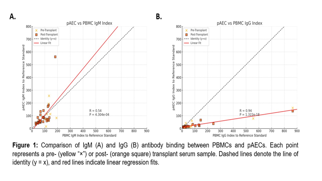
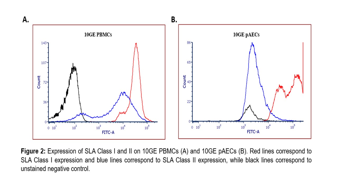
Conclusion: These findings suggest that IgM-targeted epitopes are consistently preserved across PBMCs and pAECs, while IgG-targeted epitopes may be absent on pAECs or selectively lost during endothelial culture. The diminished IgG binding observed on pAECs, combined with reduced SLA-II expression, suggests that cell surface marker expression may influence the sensitivity of pAEC-based assays for detecting IgG-dependent xeno-reactivity. These results highlight the importance of including PBMC-based assays alongside pAEC-based assays for comprehensive antibody testing in upcoming kidney xenotransplant trials. Accrual of these data will be crucial not only for immunologic risk assessment and post-transplant monitoring but also for discovery of clinically relevant xenoantigens.
This study was supported by a grant from United Therapeutics Corporation.
[1] Immunologic screening
[2] Xenocrossmatch
[3] Xeno-antibody monitoring after transplantation
[4] porcine aortic endothelial cells
[5] porcine peripheral blood mononuclear cells