Pig kidney xenograft size after pig-to-human transplantation - GGTA1 KO Thymokidney vs 10 GE Xenokidney
Tal Eitan1, Imad Aljabban1, Jacqueline Kim1, Ian Jaffe1, Aprajita Mattoo1, Vasishta Tatapudi1, David Ayares2, Elaina Weldon1, Karen Khalil1, Jayme Locke2, Adam Griesemer1, Robert A Montgomery1, Jeffrey Stern1.
1NYU Langone Transplant Institute, New York, NY, United States; 2United Therapeutics Corp., Durham, NC, United States
Introduction: As xenotransplantation studies in humans evolve in both decedents and living recipients, different genetic edits and immunosuppressive regimens are being optimized to overcome immunologic barriers. We compared post-transplant pig xenograft sizes of GGTA1 KO Thymokidneys, in a decedent and living recipient, and a 10 GE Xenokidney in a living recipient.
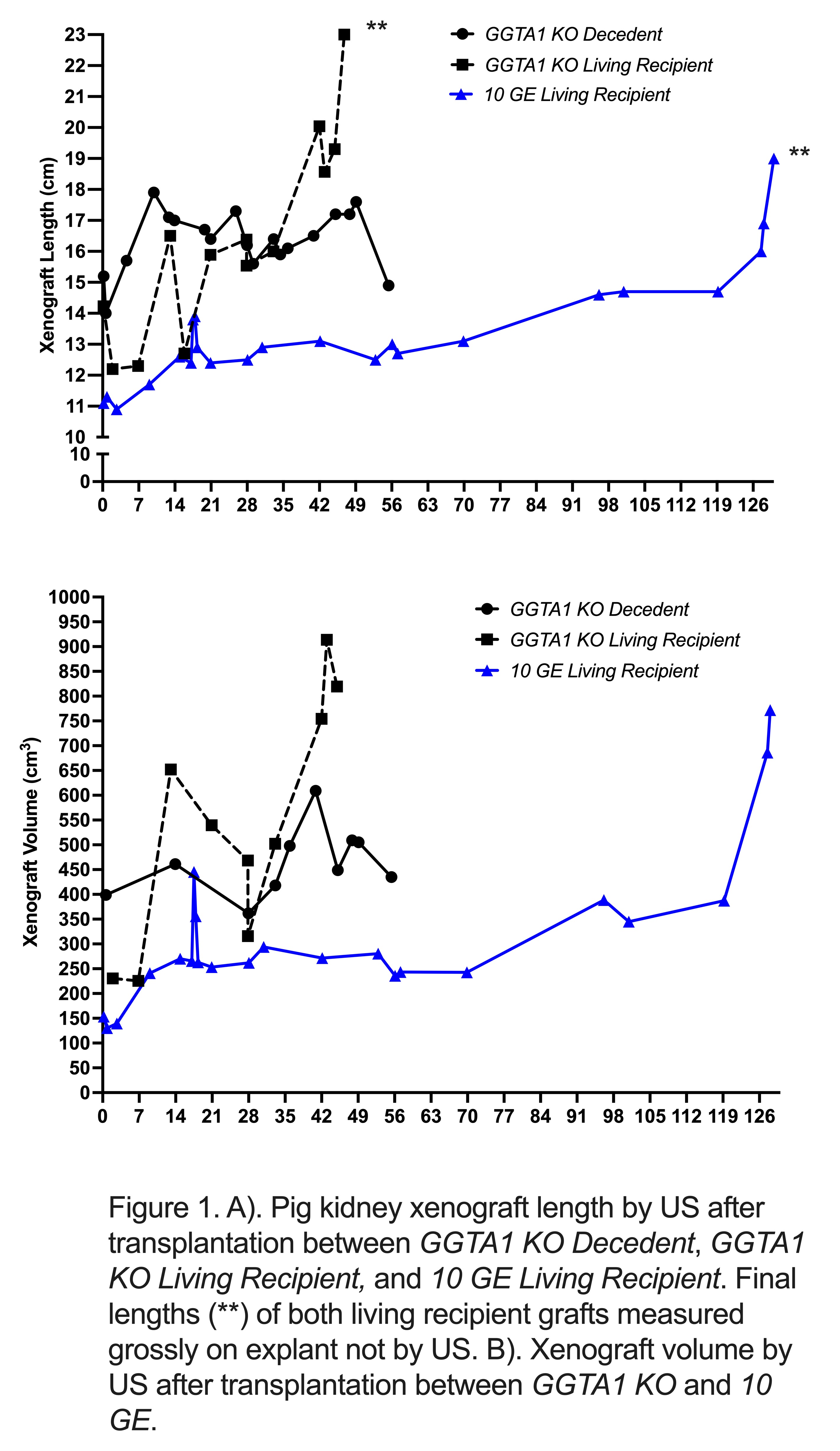
Methods: A 57-year-old male brain-dead human decedent underwent bilateral nephrectomy and xenotransplant of a 203 g GGTA1 KO Thymokidney. Source pig was a 80.kg, 6.6-month-old landrace swine. Postoperatively, this decedent was somatically supported for 61 days before elective termination. A 54-year-old female living human with multiple co-morbidities including end-stage heart and kidney disease was determined to not be a heart-kidney transplant candidate and underwent xenotransplant with a 268 g GGTA1 KO Thymokidney 8 days after implantation of a HeartMate 3 LVAD. Source pig was a 200 kg, 11-month-old landrace swine. A highly sensitized living human underwent transplant of a 240 g 10 GE Xenokidney, including knockouts of 3 known immunogenic carbohydrates and the porcine growth hormone receptor and human transgene expression of proteins responsible for inhibiting human complement and coagulation pathways. Source pig was a 99.3 kg, 15-month-old landrace swine. Ultrasound (US) measurements were collected during per-protocol and for-cause exams using Seimens Acuson S3000. A combination of direct volumetric scanning and calculation of length, width, and depth from US exams was used.
Results: In the GGTA1 KO Thymokidney in the decedent, by POD 10, graft length increased by 2.7 cm (Figure 1A). By POD 14, graft volume increased by 60 cm3. Volume increased by 150 cm3 during an episode of rejection that began on POD 33. Volume decreased by 320 cm3 with treatment. In the GGTA1 KO Thymokidney in the living recipient, by POD 13, graft length increased by 4.3 cm and volume increased by 420 cm3. From POD 33 to 45, renal function declined without evidence of immunologic rejection and on POD 45 volume had increased by 167 cm3 and length had increased by 6.5 cm vs POD 13. In the 10 GE Xenokidney, by POD 9, graft length increased by 0.6 cm and volume increased by 110.7 cm3. During an episode of rejection that started on POD 17, volume increased by 53.3 cm3 and length increased by 2.1 cm. By POD 57, volume returned to baseline with treatment (Figure 1B). A second episode of rejection in the last week of the study yielded volume increase of 530 cm3 and length increase of 7 cm.
Conclusion: All three xenografts experienced expected early size increases, likely due to hyperfiltration. None of the xenografts had clinically significant volume changes. Volume changes may be more suggestive of immune factors than dysregulated growth as volumes decreased after rejection treatment. The volume increase in the GGTA1 KO Thymokidney in the living recipient was likely related to edema from hemorrhagic necrosis.
Tal Eitan was supported by the National Cancer Institute, National Institutes of Health, through Grant Award Number T32 CA193111. . Work funded in part by Lung Biotechnology, a wholly owned subsidiary of United Therapeutics Corporation..
[1] Xenograft size, GGTA1 KO Thymokidney, 10 GE Xenokidney